|
a |
 b |
|
|
1A
|
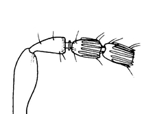
Antennal pedicel
longer than first funicle segment; first funicle segment with one
transverse row of sensilla (may be irregular) or with only a single
sensillum visible in dorsal view.
|
2 |
|
1B
|
Antennal pedicel shorter than or
equivalent to first funicle segment; first funicle segment with more than one
row of sensilla, which may form definite or integrated rows, but clearly not one
row. |
10 |
|
2A
|
Basal portion of
stigmal vein almost perpendicular to the wing margin; wing club-shaped,
its length less than two and half times the width |
3 |
|
2B
|
Basal portion of
stigmal vein at an acute angle to the wing margin; wing more elongate,
its length more than two and half times the width
|
8 |
|

a |
b
|
c
|
|
3A
|
Fore wing clear with
few microtrichia, almost glabrous |
4 |
|
3B
|
Fore wing with one
or two dark patches, many microtrichia |
5 |
|
4A
|
All of body metallic
greenish-black, ovipositor barely projecting beyond the end of the
metasoma.
|
D. bizarrea |
|
4B
|
Only head metallic
greenish-black – mesosoma and metasoma yellowish-brown; ovipositor
projecting well beyond end of metasoma. |
D. falcata
|
|

a |
|
|
|
5A
|
Two dark patches on
the fore wing, one below the stigmal vein and adjacent to the marginal
vein the other above the stigmal vein. |
D. bicolor
|
|

a |

b |
c
|
|
5B
|
One small or very
large dark patch on the fore wing, below the stigmal vein and adjacent
to the marginal vein. |
6 |
|
6A
|
Hypopygium not
projecting beyond the end of the metasoma (fig a); mesosoma metallic
greenish-black, except for yellow propodeum (fig b); dark patch large, extending
across the entire width of the wings (fig c). |
D. yangi
|
|
a |
b |
 c
|
|
6B
|
Hypopygium
projecting beyond the end of the metasoma; mesosoma yellow; dark patch
on forewing small, restricted to area adjacent to marginal vein. |
7 |
|
7A
|
Head with the vertex
raised towards the ocelli (fig a); clypeal margin with small cone-like central
protuberance (fig a). Mandible bidentate tricuspidate and three times as long as
wide (fig b). Antennal insertion clearly above ventral margin of the eye
(fig c). |
D. latipennis |
|
7B
|
Head with vertex
truncate (fig a), clypeal margin more indented, with a central (but not cone
like) protuberance. The sides of the protuberance smooth and leading up
to a fairly sharp central point (fig a). Mandible bidentate bicuspidate, with a
slight protuberance on the inner margin of the subapical tooth; four
times as long as wide (fig b). Antennal insertion just above ventral margin of
the eye (fig c). |
D. laticeps |
|
8A
|
Fore wing almost
four times as long as wide (figs a & b). Clypeal margin concave with two slight
projections on either side of a shallow central concavity (fig c).
|
D. macroptera
|
|
8B
|
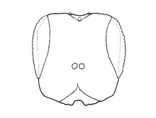
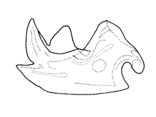
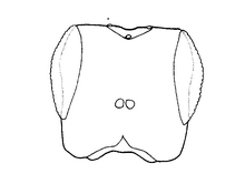
Fore wing less than
three and a half times as long as wide (fig a). Clypeal margin either slightly
protruding with a central U-shaped concavity (fig b), or broadly concave with a
slight central convexity (fig c). |
9 |
|
 a |
 b
|
|
|
9A
|
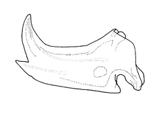
Clypeal margin
slightly protruding with a central U-shaped concavity (head more rounded
in shape) (fig a). Mandible bidentate tricuspidate, length approximately two
and a half times the width (fig b). |
D. alleni |
|
 a |
b |
|
|
9B
|
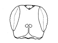
Clypeal margin
broadly concave with a very slight central convexity (head more square
in shape) (fig a). Mandible bidentate bicuspidate (the subapical tooth may have
a small protuberance on the inner margin), length almost four times the
width (fig b). |
D. wiebesi |
|
a |
|
|
|
10A
|
Head yellow in
colour; clypeal margin with three slight protuberances, one in the
centre and one on either side of the central protuberance (the
protuberances being broadly separated) (fig a). |
D. pallidiceps
|
|
10B
|
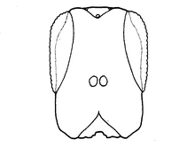
Head metallic
greenish-black in colour, clypeal margin either with a central concavity
(fig a)
or central truncate protuberance (figs b & c). |
11 |
|
 a |
 b
|
|
|
11A
|
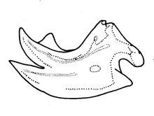
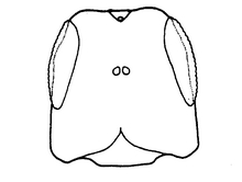
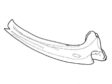
Clypeal margin with
a central concavity (fig a). Mandible tridentate and short, twice as long as
wide (fig b). |
12 |
|
 a |
b |
|
|
11B
|
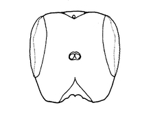
Clypeal margin with
a central truncate protuberance (fig a). Mandible bidentate and elongate, length
more than six times the width (fig b). |
13 |
|
 a |
|
|
|
12A
|
Central concavity on
clypeal margin scalloped with sharp medial point. Head wider than long
(1.09:1). Shortest distance between compound eyes slightly greater
(1.1X) than eye length (fig a). |
D.
philippinensis |
|
 a |
|
|
|
12B
|
Clypeal margin with
a central U-shaped concavity that has stepped sides. Head clearly longer
than broad (1.15:1). Shortest distance between compound eyes much less
(0.68X) than eye length (fig a). |
D. longiceps |
|
 a |
 b
|
|
|
13A
|
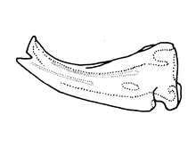
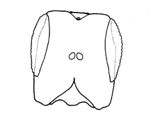
Mandible extremely
long and narrow, the length more than nine times the width (fig a). Antennal
insertion clearly closer to the vertex than to the clypeal margin.
Clypeal margin with a broad truncate protuberance (width approximately
one third the width of the head); cheeks diverging towards the ventral
margin (fig b). |
D. tumidigena
|
|
 a |
b |
|
|
13B
|
Mandible length six
to seven times longer than wide (fig a). Antennal insertion approximately mid
way between the vertex and the clypeal margin. Clypeal margin with a
smaller truncate protuberance (width approximately one seventh the width
of the head); cheeks sub-parallel, slightly converging towards the
ventral margin (fig b). |
D. retakensis |
|
References |
Gardiner,
A.J. & Compton, S.G. 1987.
New species of the fig wasp genus Diaziella
(Hymenoptera,
Chalcidoidea, Sycoecinae). Tijdschrift
voor Entomologie 130:129–140.
Grandi,
G. 1928. Un nuovo genre quattro nuove specie di imenotteri sicofili di Sumatra. Bollettino
del Laboratorio di Entomologia del R.Istituto Superiore Agrario di Bologna
1: 71-89.
van Noort, S., Peng, Y.Q. & Rasplus, J.Y. 2006.
First record of the fig wasp genus Diaziella Grandi
(Hymenoptera: Chalcidoidea: Pteromalidae: Sycoecinae) from the Asian mainland
with description of two new species from China. Zootaxa.
Wiebes,
J.T. 1974. The fig wasp genus Diaziella (Hymenoptera, Chalcidoidea,
Torymidae, Sycoecini). Proceedings Koninklijke Nederlandse Akademie van
Wetenschappen, Amsterdam (C) 77: 295-300.
|
Credits
|
Photographs © Simon van Noort (Iziko Museums of South Africa); illustrations by Alan
Gardiner.
|
|